
The Ankle (Human Ankle Anatomy) Muscles, Bones, Ligaments, All you need to know about
A general understanding of the anatomy and the working of the human ankle joint is very important since it is one of the most commonly and easily injured and sprained joints in the body. Any problem in the ankle joint can be debilitating since it makes walking on the affected leg a nightmare.
Anatomy of the Human Ankle:
Bones:
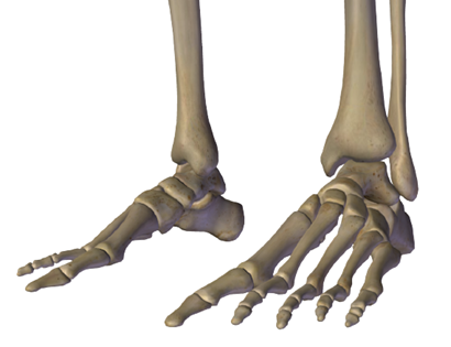
The ankle joint in the lower limb is made up of three bones:
The thick shin bone of the lower leg, also known as the tibia, the thinner bone running next to the shin bone known as the fibula, and the foot bone that sits above the heel bone, connecting to the tibia and fibula, is known as the talus.
The Bony Prominences:
The bony prominences (or protrusions) which are felt and seen on the ankle are:
The medial malleolus, which is felt on the inside of your ankle and is part of the tibia's base
The posterior malleolus, which is felt on the back of your ankle is also part of the tibia's base
The lateral malleolus which is felt on the outside of your ankle is the lower end of the fibula
Articulation:
The upper articular surface is formed by:
(i) The lower end of the tibia including the medial malleolus,
(ii) the lateral malleolus of the fibula, and
(iii) the inferior transverse tibiofibular ligament.
These structures form a deep socket.
The inferior articular surface of the ankle joint is formed by articular areas on the upper, medial and lateral aspects of the talus bone and it fits into this deep socket.
Movement and Muscles Involved at the Ankle Joint:
The ankle joint is anatomically classified as a hinge joint, this is because its movement is permitted in one plane, just like the movement of a hinge.
The movement produced at the ankle joint are;
- Plantarflexion:
It is the movement in which the top of your foot points away from your leg.
It is produced by the muscles in the posterior compartment of your leg (the calf muscles which include the gastrocnemius, soleus, plantaris, and posterior tibialis muscles).
- Dorsiflexion:
It is the backward bending and contracting of your foot. It is produced by the muscles of the anterior compartment of the leg namely the tibialis anterior, extensor hallucis longus, and extensor digitorum longus muscles.
Contrary to the popular belief, the ankle joint does not move side to side, rather movement is produced by the subtalar joint, which sits below the ankle joint.
During walking, the plantarflexors help raise the foot from the ground while the dorsiflexors help in planting the foot on the ground. This to and fro motion of the ankle joint is what enables us to walk.
| Movement | Primary Muscles | Accessory Muscles |
| Plantarflexion | 1. Gastrocnemius 2. Soleus | 1. Plantaris 2. Tibialis posterior 3. Flexor hallucis longus 4. Flexor digitorum longus |
| Dorsiflexion | Tibialis Anterior | 1. Extensor digitorum longus 2. Extensor hallucis longus 3. Peroneus tertius |
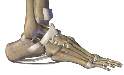
Ligaments of the Ankle:
Ligaments are cord-like bands of tissues that connect bones with each other. The ankle ligament connects the foot bones with the bones of the lower leg. They help in stabilizing the joint and prevent the joint from twisting, collapsing, or folding. Any tear or overstretching of these ligaments is what is generally known as an ankle sprain.
The joint is supported by:
(i) Fibrous capsule,
(ii) a medial (deltoid) ligament
(iii) a lateral ligament.
- Fibrous Capsule:
It surrounds the ankle joint. It is thin anteriorly and posteriorly which allows for the hinge movement, while laterally it is thickened and is supported by strong collateral ligaments.
The fibrous capsule is lined by a synovial membrane.
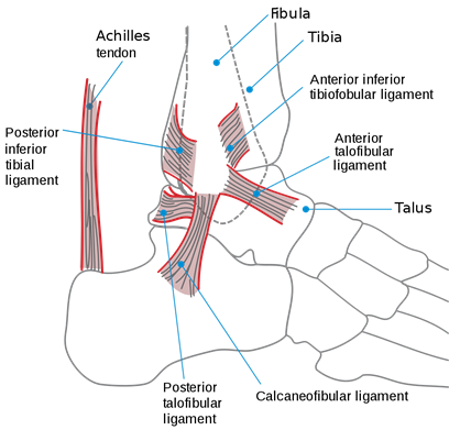
- Deltoid or Medial Ligament:
It is a strong triangular ligament on the inside (medial side) of the ankle. It is crossed by tendons of the tibialis posterior and flexor digitorum longus .
- Lateral Ligament: This ligament consists of three bands of ligaments, which are;
1. The anterior talofibular ligament which connects the anterior of the lateral malleolus to the neck of the talus, as shown in the figure below.
2. The posterior talofibular ligament which passes from the lower part of the malleolar fossa of the fibula and attaches to the lateral tubercle of the talus.
3. The calcaneofibular ligament, which is a long rounded cord passing from the notch on the lower border of the lateral malleolus and connecting to the tubercle on the lateral surface of the calcaneum.
Blood Supply of the Ankle Joint:
The ankle joint is supplied by the tibial arteries (the anterior and posterior), and peroneal arteries.
Nerve Supply of the Ankle Joint:
The innervation to the ankle joint is provided by tibial, superficial fibular and deep fibular nerves.
Ankle Conditions and Disorders:
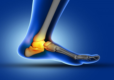
Some of the medical conditions involving the ankle are;
| Ankle Sprain: | It is an ankle injury that occurs when the ligaments of the ankle stretch or twist beyond their normal limits which results in tears of the ligaments. It is the most common type of ankle injury accounting for 2 million injuries per year. Sprains usually occur due to excessive abduction of the subtalar joint (it might involve some fibers of the deltoid ligament as well). Forced Plantarflexion of the ankle joint causes sprain by tearing the anterior fibers of the fibrous capsule. Though most of the ankle sprains are sports-related, others are related to causes like wearing high heels. |
| Dislocations of the ankle: | Because of the stability provided the ankle ligaments and joint capsule, dislocation of the ankle occurs very rarely. When it occurs it is usually along with ankle fractures. |
| Ankle Fracture: | It is a condition in which a fracture in any of the 3 bones that make up the ankle joint (the tibia, fibula, and talus) occurs. Most commonly, the bones of the lower leg (tibia and fibula) are the ones fractured. |
| Ankle Arthritis: | Arthritis is an inflammation and degeneration of any joint in the body. It can occur in the ankle joint as well. |
| Rheumatoid Arthritis: | It is a form of arthritis in which your body's immune system attacks your joint and it can affect your ankle joint, along with other joints in your body |
| Gout: | It is a form of arthritis in which uric acid crystal deposits in the joint and lead to its inflammation and degeneration. The ankle joint can be affected just like other joints of the body. |
| Psoriatic Arthritis: | Psoriasis is a skin condition that causes red, itchy scaly patches on the skin. It is usually associated with arthritis of the joint as well, which is known as psoriatic arthritis. |
| Septic Arthritis: | Any bacterial infection can occur in the ankle joint and lead to painful arthritis known as septic arthritis. |
| Pott’s Fracture: | It is the fracture of one or more of the prominences of the ankle (the malleoli). It usually occurs in plays like basketballs while landing from a jump, or when rolling an ankle. |
| Peroneal tendon tears : | The peroneal tendons are two parallel tendons on the outer ankle bone that function in protecting the ankle from sprains. In sports involving prolonged ankle movements like soccer, the peroneal tendon can tear. |
| Ankle impingement: | It is the impingement of the nerves that supply the ankle joint. It can lead to chronic anterior ankle pain and swelling around the ankle. |
Ankle Tests:
- Physical Examination:
In this test, the physician examines the ankle joint and diagnosis whether the pain and swelling in the ankle are because of a sprain, fracture, or any other reason.
- Ankle X-ray:
A simple X-ray can help the physician visualize the fracture, sprain, or tear.
- Stress test
The physician puts the ankle in a stressful position, in line with the ligament tear, and scans the ankle through an x-ray to diagnose the ankle condition.
- MRI Scan:
MRI scans, just like X-rays, can be used to identify ankle conditions. The MRI scanner creates high-resolution images of the ankle.
Ankle Condition Treatments:
- Pain medications:
Most painkillers can help soothe ankle pain. Some may be available over the counter while others would require a physician's prescription. Some common painkillers include:
- Aspirin
- Paracetamol
- Tylenol
- RICE therapy
RICE therapy has been the most famous initial therapy for ankle pain. RICE stands for Rest, Ice, Compression, and Elevation. It involves allowing the ankle to rest, applying ice packs on it, compressing it using athletic bands, and raising the leg to elevate the ankle and relieve the ankle pain.
- Casting (Ankle Immobilization)
Most ankle fractures require the ankles to be immobilized using a cast to make sure of the proper recovery from the fracture. Some physicians would cast the ankle up even in cases of an ankle sprain.
- Surgical Treatments
In cases of severe and complicated ankle conditions, the ankle usually ends up requiring surgical treatment.