
Feet Human Feet Anatomy, Muscles, Bones, Ligaments All you need to know about
The feet are the lowest and the last part of the human body that is responsible for keeping our balance and helping us walk and run. They are external body parts that are made up of muscle, bones, ligaments, tendons, and joints.
The feet's shape is also responsible for the natural balance of the body when standing as all the weight of your body is centered at your feet.
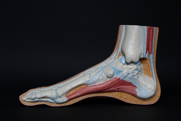
They are made up of several small and large bones, calcaneus being the largest of all. The calcaneus is commonly known as the heel bone.
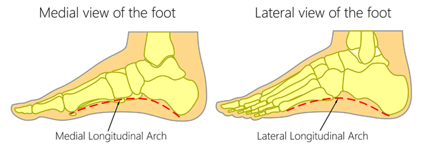
The foot is arched medially and laterally. The arches help it function properly. Some people have flat foot due to improper arches. This condition can be very troublesome.
The foot receives a widespread blood supply through arteries and their sub-branches and has venous and lymphatic drainage systems. The nerve supply of the foot is also very extensive.
Sections
The feet are categorized into three sections
| Forefoot | Midfoot | Hindfoot |
| It includes the five phalanges (toes) and the five metatarsal bones. | The three bones cuneiform bone, cuboid bone, and navicular bone form a pyramid collection that forms the arches of the feet. | It includes the remaining portion of the foot, the heel, and the ankle. The talus bone forms the ankle and supports the bones of the leg (tibia and fibula). |
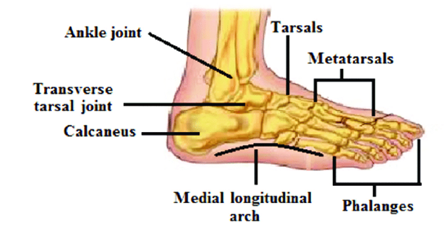
Bones of the foot
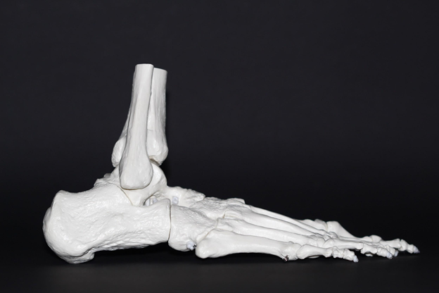
There are 26 bones in each foot. The bones of the foot are named as:
- Tarsal Bones
- Metatarsals
- Phalanges
These bones make the toes and feet.
Connecting Bones
The other bones of the foot create the ankle. The connecting bones are
- Tibia and Fibula
- Talus
- Cuneiforms
- Cuboid
- Navicular
Tarsal Bones
The tarsal bones are a set of seven bones. The tarsal bones are irregular. They are located in the ankle area.
The tarsal bones are subdivided into three groups
- Proximal Group (Hindfoot)
- Intermediate Group (Midfoot)
- Distal Group (Midfoot)
Proximal Group
The proximal part of the heel and ankle is formed by the proximal tarsal bones known as the Talus and the Calcaneus.
- Talus:
The talus is the superior proximal tarsal bone. It is responsible for transmitting the bodyweight to the foot.
It articulates with other joints for movements.
- Calcaneus:
It is the longest tarsal bone. It articulates (make joints) with the talus and cuboid bones. It takes the weight of the body while walking.
Intermediate Group
The intermediate group contains one bone only the navicular bone. The name navicular is due to its shape like a boat.
It articulates with the talus bone, cuneiform bones, and cuboid bone.
Distal Group
The cuboid and the three cuneiform bones form the distal group of tarsal bones.
Metatarsals
The metatarsals are located between tarsals and phalanges. The metatarsals comprise the forefoot.
There are four metatarsal bones. The metatarsals have the same structure.
- Convex Dorsally
- Head
- Neck
- Shaft
- Base
The metatarsal bones have three to four articulations.
Phalanges
The bones of the toes are known as phalanges. There are fourteen phalanges.
- The big toe has only two phalanges
- Proximal
- Distal
- The other toes from the second toe to the fifth toe are
- Proximal
- Middle
- Distal
Joints
There is an ankle joint that connects the foot and leg. The bones of the foot articulate with each other as well through joints. The four main groups are as well
Intertarsal Joints: As the name indicates these are the joints between tarsal bones.
Tarsometatarsal Joints: The joint between the tarsals and metatarsals.
Metatarsophalangeal Joints (MTP): Metatarsophalangeal joints are the joints between the head of metatarsals and bases of phalanges of the foot.
Interphalangeal Joints: These are the joints between the phalanges.
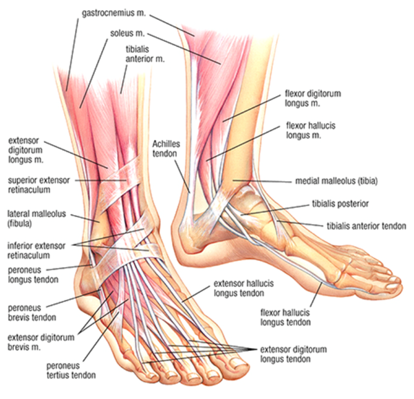
Muscles of the Foot
Muscles help in various types of movement such as eversion, inversion, and plantar flexion, and dorsiflexion. The foot muscles are grouped into plantar and dorsal groups. The plantar muscles are divided into three groups
- Lateral
- Central
- Medial
Dorsal Foot Muscles
The dorsal muscle has two muscles in the dorsum of the foot. The muscles extend to the toes. They are:
- Extensor Digitorum Brevis:
They are involved in toe extension and distal interphalangeal joints.
- Extensor Hallucis Brevis:
The function of this muscle is involved in the movement of metatarsophalangeal joints. It is involved in toe extension.
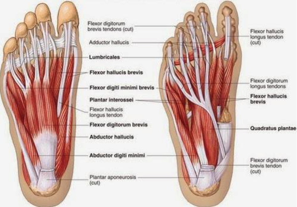
Plantar Foot Muscles
The plantar muscle is divided by the deep fascia of the foot into three groups.
- Lateral plantar muscles
- Central planar muscles
- Medial plantar muscles
Lateral Plantar Muscles
There are three lateral plantar muscles. They act upon the fifth toe of the foot. They are named as
- Abductor Digiti Minimi
It supports the longitudinal arch of the foot and toe flexion. It also helps in toe abduction. It assists in the movement of metatarsophalangeal joints.
- Flexor Digiti Minimi Brevis
It assists in toe flexion.
- Opponens Digiti Minimi
It helps you in the toe abduction and toe flexion.
Central Plantar Muscles
They are further divided into five muscles. They act upon the four lateral toes. They are:
- Flexor Digitorum Brevis
Like abductor digiti minimi, It also supports the longitudinal arch of the foot.
- Quadratus Plantae
It also helps in toe flexion. It helps in the movement of metatarsophalangeal joints.
- Lumbricals
It helps in the movement of interphalangeal joints and metatarsophalangeal joints. It helps in toe adduction and toe extension.
- Plantar Interossel
It is involved in toe flexion and toe adduction. And like lubricants, it also assists the same two joints
- Dorsal Interossal
It is involved in toe flexion and toe abduction. It assists the metatarsophalangeal and interphalangeal joints.
Medial Plantar Muscles
It acts upon the great toe. It has three muscles. They are named as follows:
- Abductor Hallucis Muscle
It supports the longitudinal arch of the foot.
- Adductor Hallucis Muscle
It supports the longitudinal and transverse arches of the foot.
- Flexor Hallucis Brevis Muscle
It supports the longitudinal arches of the foot and toe flexion.
Tendon & Sheaths
A tendon sheath wraps around a tendon and prevents friction and helps in movement. The sheath secretes synovial fluid which is moist and lubricates the tendon.
The retinacula (fibrous bands) make a shaft around the tendon.
- Anterior Sheaths
- Posterior Sheaths
Anterior Sheath
Anterior Sheaths cover the four tendons of the foot. There are three anterior sheaths
First Sheath: This sheath covers the tibialis anterior tendon. It divides into two limbs at the inferior extensor retinaculum.
Second Sheath: The common sheath for the fibularis Tertius and extensor digitorum longus muscle.
Third Sheath: The third sheath covers the extensor hallucis longus tendon.
Posterior Sheaths
There are four posterior sheaths.
First Sheath: This sheath encompasses the tibialis posterior tendons.
Second Sheath: This sheath wraps the Flexor hallucis longus tendon.
Third Sheath: This sheath shuts in the flexor digitorum longus.
Fourth Sheath: This sheath encloses the muscles fibularis longus and fibularis brevis muscle.
The Arches
The foot has three arches. The arches are formed by bones and supported by muscles and ligaments. The foot arches facilitate walking and running. They help the foot in bearing the whole body weight.
Medial Arch: The medial arch is formed by the calcaneus, talus, navicular and cuneiforms, and metatarsal bones.
It is supported by muscles such as intrinsic foot muscles and flexor hallucis. The plantar and medial ligament supports the medial arch. It has plantar aponeurosis.
Lateral Arch: It is flatter than the medial arch. It is formed by the 4th and 5th metatarsal bones, calcaneus bone, and cuboid bone.
It is supported by fibularis longus and flexor digitorum longus. The plantar ligament supports it. It also has plantar aponeurosis.
Transverse Arch: It is in the coronal plane of the foot. The transverse arch is constructed by metatarsal bones, the cuboid, and the three cuneiform bones.
The muscles that support the transverse arch are the fibularis longus and tibialis posterior. The plantar and deep metatarsal ligament support it. It has plantar aponeurosis.
Blood Supply
The blood is supplied to the foot via two arteries.
Arterial Supply
The blood is supplied to the foot via two arteries
- Dorsalis Pedis
- Posterior Tibial
Venous Drainage
The venous drainage of the foot is by the dorsal venous arch. The veins are named as follows
- Anterior Tibial vein
- Posterior tibial vein
- Posterior fibular vein
- Medial Malleolus
- Popliteal vein
- Adductor canal
Lymphatic Drainage
The lymph of the lower limb is carried by two groups
Superficial Lymphatic Vessel
The superficial lymphatic vessels are further divided into two groups
- Medial Vessels
It originates from the dorsal surface of the foot. It passes through the medial condyle of the femur. Ut drains into
- Subinguinal groups of lymph nodes.
- Lateral Vessels
They arise from the lateral surface of the foot. They either drain into popliteal nodes or join the medial lymphatic vessels.
Deep Lymphatic Vessel
The deep lymphatic vessels accompany the deep arteries of the lower leg. They are named as
- Anterior Tibial
- Posterior Tibial
- Peroneal
Common Diseases
The common foot problems include
- Athletes foot
- Foot pain
- Stress Fracture
- Diabetic Foot Ulcer
Precautionary Measures
To take care of your feet please follow the following steps
- Easy Medicated Shoes
- Soft Soul
- Exercise of feet
- Give proper rest to your feet