
Shoulder Joint Anatomy, Pictures, Structures Conditions and More
The shoulder joint is the combination of several joints that combine with tendons and muscles to allow a wide range of motion in the arm, such as swinging the bat, climbing, swimming, and catching a ball, etc. In the human body, the shoulder is one of the largest and most complex joints.
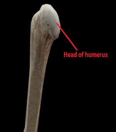
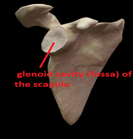
A ball-and-socket joint forms when the upper arm bone (humerus) meets the shoulder blade (scapula). As one of the largest and most complex joints in the body, the shoulder is also one of the most vulnerable.
In this article, we will study in detail the basic anatomy of the shoulder along with its functions and some of the common diseases of this region.
Shoulder joint:
The glenohumeral joint is another name for the shoulder joint.It is the ball and type of synovial joint that permits a wide range of movements. However, its mobility makes the shoulder joint relatively unstable.
Articulation:
It forms by articulating the humerus head laterally with the glenoid cavity (fossa) of the scapula medially. Because of this, the joint is also given an alternative name known as the glenohumeral joint.
The articulating surfaces of most synovial joints are covered in hyaline cartilage. Shoulder joints are no different.Due to the giant head of the humerus than the glenoid fossa, the joint can move more freely, but it is inherently unstable.
The surface disproportion is reduced By deepening the glenoid fossa with a cartilage rim called the glenoid labrum.
Joint capsule:
The shoulder joint is surrounded by the fibrous joint capsule. The joint capsule is attached medially to the margin of the glenoid cavity and laterally to the anatomical neck of the humerus.
- The part of the capsule lines the coracoid process so that the fibrous layer of the capsule surrounds the proximal attachment of the long head of the biceps brachii. There are two apertures in the joint capsule:
- An opening between the tubercles of the humerus to allow the passage of the tendon of the long head of the biceps brachii.
- An opening situated anteriorly, inferior to the coracoid, the process that allows communication between the subtendinous bursa of the subscapularis and the synovial cavity of the joint.
- The weakest area lies in the inferior part of the joint capsule, the only part not reinforced by the rotator cuff muscles. The capsule is particularly lax here and lies in folds when the arm is adducted; however, when the arm is abducted, it becomes taut.
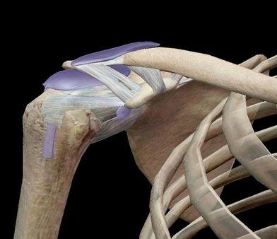
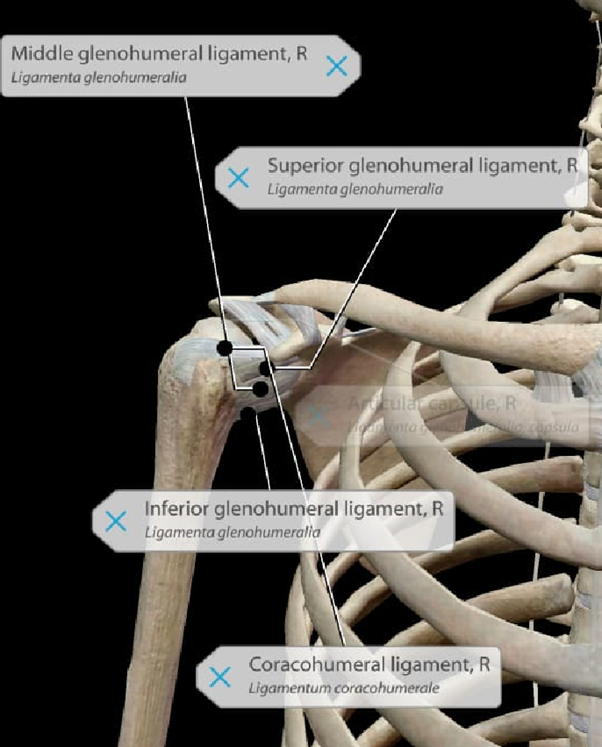
Ligaments of the Shoulder:
Following are some of the major ligaments of the shoulder joints:
| Glenohumeral ligaments | These ligaments connect the humerus to the glenoid fossa, forming the joint capsule. Besides holding the shoulder in place and preventing it from dislocating anteriorly, they also provide principal stability to the shoulder. The anterior aspects of the joint are stabilized by these structures. So these ligaments are very important for the shoulder joints. |
| coracohumeral ligament | It connects the coracoid process to the greater tubercle of the humerus. It provides support to the superior portion of the joint capsule. |
| Transverse humeral ligament | It extends between the humerus's two tubercles. In the intertubercular groove, this ligament supports the long head of the biceps. |
| Caraco–clavicular ligament | Lying between the clavicle and the coracoid process of the scapula, Caraco–clavicular ligament is composed of the trapezoid and conoid ligaments. Their function is to align the clavicle concerning the scapula, as they act in conjunction with the acromioclavicular ligament. In an acromioclavicular joint (ACJ) injury, these ligaments are strong, but large forces can rupture them. A severe ACJ injury may require surgery to repair the coracoclavicular ligaments. |
Factors that contribute to the mobility and stability of shoulder joint:
Despite its mobility, the shoulder joint is one of the most unstable in the body. We will investigate factors that allow movement, as well as those that contribute to joint structure:
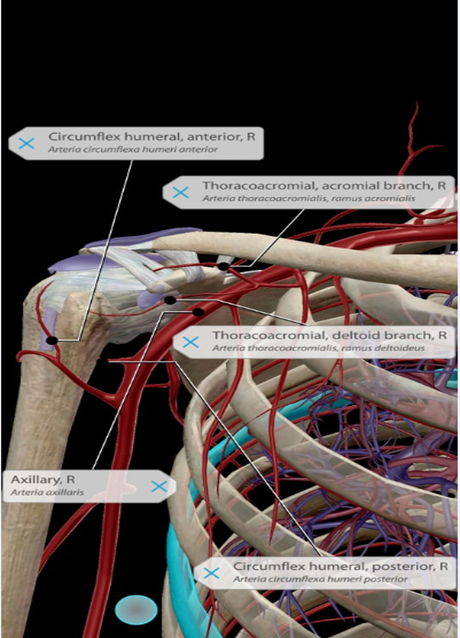
Blood supply of Shoulder:
The shoulder is supplied with blood by four main arteries:
| The subclavian artery | Under the collarbone is the subclavian artery. Through it, blood is supplied to the shoulder. |
| The axillary artery | In the armpit is the axillary artery. As a continuation of the subclavian artery, the axillary artery provides blood to the upper arm. |
| The thoracoacromial artery | At the top of the shoulder, the thoracoacromial artery arises from the axillary artery. From this artery, four branches branch out into the shoulder and upper chest region. |
| The brachial artery | This artery runs down the arm. It is an extension of the axillary artery. Blood is supplied to the muscles and bones of the shoulder by branches of the brachial artery. |
Nerve Supply of Shoulder:
All the nerves supplying the upper limb pass through the axilla(the armpit) just under the shoulder joint. They are derived from the plexus of the nerves known as the Brachial Plexus before dividing into individual nerves. Signals from the brain are sent to these nerves, which move the arm's muscles. Sensations such as touch, pain, pressure, and temperature are transmitted by the nerves to the brain.
Many nerves make up the Brachial Plexus, which are responsible for supplying the arm with the ability to function. Some of the common nerves are as follows:
- The axillary nerve supplies the Deltoid muscle.
- Long Thoracic nerve supplies Serratus Anterior muscle.
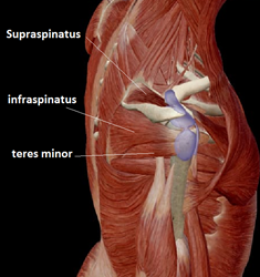
- The suprascapular nerve supplies supraspinatus and infraspinatus muscles.
- The musculocutaneous nerve supplies the Biceps muscle.
- The radial nerve
- The ulnar nerve
- The musculocutaneous nerve
- The median nerve
Movements of Shoulder Joint:
As the shoulder joint is a ball and socket synovial joint, it allows a wide range of movement permitted:
| Type of movement | Explanation | Muscles involved |
| Extension | Movement of upper limb backwards in the sagittal plane | posterior deltoidlatissimus dorsi teres major. |
| Flexion | Movement of upper limb forwards in the sagittal plane | pectoralis majoranterior deltoid CoracobrachialisBiceps brachii |
| Abduction | Movement of an upper limb away from the midline in the coronal plane | Supraspinatus (0-15 degrees)Deltoid (15-90 degrees)trapezius and serratus anterior (past 90 degrees) |
| Adduction | Movement of an upper limb towards the midline in the coronal plane | pectoralis majorlatissimus dorsiteres major. |
| Internal rotation | rotation of the upper limb towards the midline, so that the thumb is pointing medially | subscapularis pectoralis major latissimus dorsiteres majoranterior deltoid |
| External rotation | Rotation of the upper limb so that the thumb points laterally away from the midline | infraspinatusteres minor |
| Circumduction | Combination of all the movements produced at the shoulder joint | All the muscles of the shoulder joint are mentioned above. |
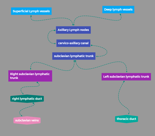
Lymphatic drainage of Shoulder:
Lymphatic vessels:
Lymph nodes:
The axilla contains the majority of lymph nodes of the upper extremity. These lymph nodes are grouped anatomically into five groups:
Through the cervico-axillary canal, lymphatic vessels from apical axillary nodes converge to form the subclavian lymphatic trunk. The right subclavian trunk forms the right lymphatic duct and enters directly into the right venous angle (joint of the internal jugular and subclavian veins). Subclavian lymphatic drainage drains directly into the thoracic duct on the left side.
Muscles of Shoulder:
Following are the muscles of the shoulder regions:
- Rotator cuff muscles include:
- Supraspinatus
- infraspinatus
- teres minor
- Subscapularis
- Rhomboid minor
- Trapezius
- Deltoid muscle
- Biceps brachii
Diseases related to Shoulder:
Following are some of the common diseases of the shoulder:
| Avascular necrosis | During avascular necrosis, the blood supply to the bones is temporarily or permanently cut off. In the absence of blood supply, the bone tissue dies and collapses. The joint surface may collapse if avascular necrosis occurs near a joint. Any bone can be affected by this condition. |
| Bursitis | Bursitis refers to the inflammation of a bursa. Bursae are closed, fluid-filled sacs that function as cushioning and gliding surfaces between tissues within the body. |
| Dislocation of the shoulder: | This occurs when your upper arm bone pops out of the cup-shaped socket in your shoulder blade. As the most mobile joint in the body, the shoulder is vulnerable to dislocations. You should seek medical treatment immediately if you suspect a dislocated shoulder. |
| Frozen shoulder | Frozen shoulder, or adhesive capsulitis, is a painful condition that limits shoulder movement. This condition occurs when the strong connective tissue surrounding the shoulder joint (called the shoulder joint capsule) becomes inflamed. |
| Rheumatoid arthritis | Joint pain and damage throughout your body are symptoms of rheumatoid arthritis (RA), an autoimmune disease. RA causes joint damage on both sides of the body. If a joint in one of your arms or legs are affected, likely, the same joint will also be affected in the other arm or leg. |