
The Stomach Anatomy & All You need to know about
The stomach is a muscular organ that helps us to process food, located on the left side of the abdomen. Between the esophagus and small intestine is the stomach, an expanded part of the digestive tract. Ingested food is accumulated in this organ, which is chemically and mechanically prepared for digestion and passage into the duodenum.
It serves as a blender and reservoir for food, but its main function is enzymatic digestion. A mass of food is converted by gastric juice into a semiliquid mixture, chyme (G. juice), which passes fairly quickly into the duodenum. Empty stomachs are only slightly larger than large intestines, but they can hold up to three litres of food.
The function of the stomach:
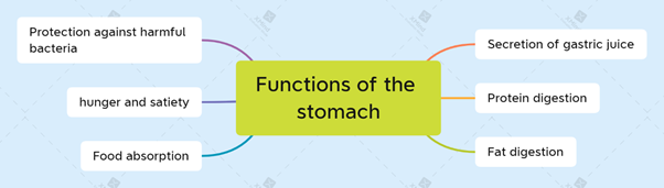
| Secretion of gastric juice | The process occurs in three stages and is mainly controlled by neuronal and hormonal factors. |
| Protein digestion | The process of digesting protein begins in the stomach. When proteins are exposed to HCl, they slowly denature (for example, actin and myosin from meat), causing structural changes and exposing the peptide bond between adjacent amino acids. |
| Fat digestion | The chief cells synthesize gastric lipase, which amplifies fat digestion in the stomach. Unlike salivary lipase, gastric lipase remains active at a wide pH range (2-7). However, it works best at a pH of 4-5 and so is most effective in the stomach. |
| Food absorption | Nutrients are not absorbed much in the stomach. It is generally believed that digested fat is not absorbed in the stomach and that only a small amount of protein digested is absorbed. However, simple sugars such as glucose, galactose, and fructose are readily absorbed, particularly when in high concentration. An average amount of water taken in by the stomach will be absorbed within 20 minutes, approximately 50% of what has been taken in. Alcoholic beverages also contain ethanol, which is rapidly absorbed across the gastric wall into the bloodstream. This illustrates how effective the stomach is at absorbing substances. |
| Hunger | Ghrelin is released by P/D1 cells of the gastric pits when the stomach is empty. The ghrelin hormone is often known as the 'hunger hormone, circulates in the bloodstream and interacts with receptors in the hypothalamus and other brain regions to cause powerful sensations of hunger. Before breakfast, lunch, and dinner, ghrelin levels are at their highest. As a result of the stomach wall stretching after eating, ghrelin is inhibited, and the sensation of hunger is diminished. |
Stomach Anatomy:
Position of the stomach: Stomach Anatomy,
| In the supine position | Often, the stomach is located in the upper right and left quadrants or in the epigastric, umbilical, and left hypochondrium and flank regions |
| In the erect position | the stomach moves inferiorly due to gravity. Individuals with asthenia (thin, weak) may have their stomach extending into the pelvis. |
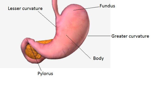
Parts of the stomach:
The stomach has four parts which are as follows:
- Cardia: This is the part surrounding the cardiac orifice (opening), also known as the upper opening or inlet of the stomach. Cardiac orifices typically lie 2–4 cm from the median plane at the level of the T11 vertebra in the supine position, posterior to the 6th left costal cartilage.
- Fundus: This is a dilated part located superiorly just left to the cardiac orifice. This is related to the left dome of the diaphragm. A cardiac notch connects the fundus and the esophagus. Any combination of gas, fluid, food, or all of them, may dilate the fundus. In the supine position, the fundus usually lies posteriorly to the left 6th rib along the MCL plane.
- Body: the region between the fundus and the pyloric antrum of the stomach.
- Pyloric part: outflow region of the stomach resembles funnel shape; its wider part, the pyloric antrum, leads into the pyloric canal, which is a narrower part. The pylorus (G., gatekeeper) is the distal, sphincteric region of the pyloric part. It is marked thickening of the circular layer of smooth muscle that controls the discharge of the stomach contents through the pyloric orifice.
Curvatures of the stomach:
The stomach also features two curvatures which are as follows:
- Lesser curvature: The lesser curvature forms the shorter concave right border of the stomach. In the most inferior part of the curvature, the angular incisure (notch) marks the junction of the body and pyloric parts of the stomach. The angular incisure is located to the left of the midline.
- Greater curvature: The greater curvatureforms the left-hand side of the stomach that is longer and more convex. It passes inferiorly to the left from the junction of the 5th intercostal space and MCL, then curves to the right, passing deep to the 9th or 10th left cartilage as it continues medially to reach the pyloric antrum. As a result of unequal lengths of the lesser curvature on the right and the greater curvature on the left, the shape of the stomach is similar to the letter J in most people.
Interior of Stomach:
During life, the smooth surface of the gastric mucosa is reddish-brown, except in the pyloric part, where it is pink. A continuous mucous layer protects its surface from the acid produced by the stomach's glands. When the gastric mucosa contracts, it forms long ridges or wrinkles called gastric folds (gastric rugae).
They are most noticeable around the pyloric part and along the greater curvature. Between the longitudinal folds along the lesser curvature, a temporary groove or furrow-like gastric canal forms during swallowing. Endoscopy and radiography can reveal this due to the attachment of gastric mucosa to the muscular layer, which lacks an oblique layer at this site, the gastric canal forms.
When the stomach is mostly empty, saliva and a small quantity of masticated food drain into the pyloric canal. As the stomach distends (fills), the gastric folds diminish and eventually disappear.
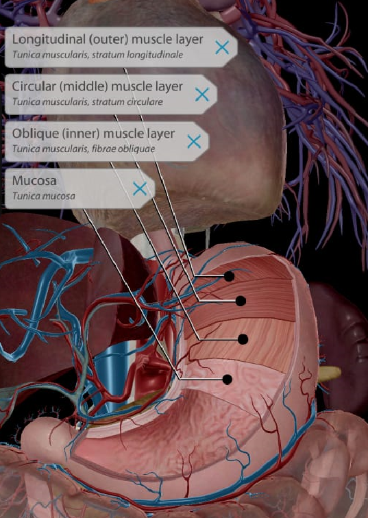
Layers of the stomach wall:
Multiple layers of tissue make up the stomach:
| The mucosa (mucous membrane) | This lining lines the inside of the stomach. The mucosa of the stomach is ridged when it is empty. As the stomach fills with food, the ridges (rugae) flatten out. |
| Submucosa | The layer below the mucosa is called the submucosa. The connective tissue that forms submucosa is composed of larger blood vessels, lymphatic vessels, nerve cells, and fibers. |
| The muscularis propria (or muscularis externa) | This layer lies beneath the submucosa. There are three layers of muscle in muscularis propria (shown in the image), which forms the main muscle of the stomach. |
| The serosa | Outside the muscularis propria is the fibrous membrane that surrounds the stomach. The stomach serosa is also called the visceral peritoneum. |
Stomach Anatomy, Sphincters of the stomach:
The contents of the stomach are contained by two smooth muscle valves or sphincters. They are the:
| Sphincter | Function |
| The cardiac or esophageal sphincter | When food is approaching, the sphincter is opened, and the food can then be swept into the stomach by rhythmic peristaltic waves. Prevent the backflow of stomach contents into the esophagus |
| Pyloric sphincter or pyloric orifice | Contributes to gastric emptying (movement of partially digested food from the stomach to the intestine). |
Stomach Anatomy, Greater and Lesser Omenta:
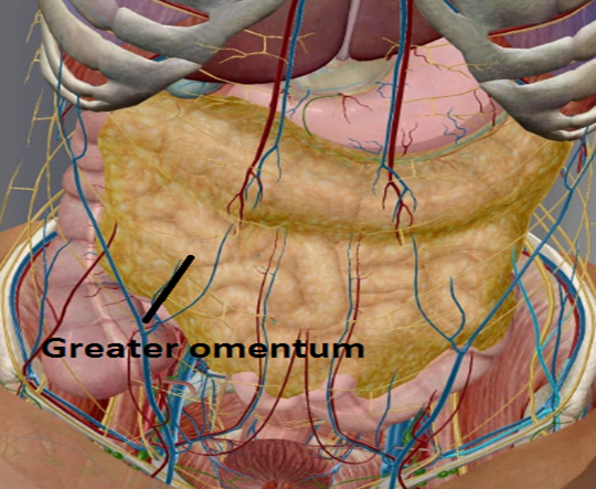
- Greater omentum: The greater omentum hangs down like a curtain from the greater curvature of the stomach, folds back upon itself, and eventually attaches to the transverse colon.
Having numerous lymph nodes and adhering to inflamed areas, plays a crucial role in gastrointestinal immunity as well as minimizing the spread of intraperitoneal infections.
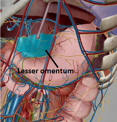
- Lesser omentum: The lesser omentum is continuous with peritoneal layers of the stomach and duodenum at one end, originating from the lesser curvature of the stomach. On the other end, it attaches to the liver. The lesser omentum attaches the stomach and duodenum to the liver.
Stomach Anatomy, Nerve supply:
| Parasympathetic nervous system | Parasympathetic innervation of the stomach originates from the anterior and posterior vagal trunks, which arise from the left and right vagus nerves (CN X), respectively. The anterior surface of the stomach and pylorus is mainly supplied by the anterior vagal trunk. The larger posterior vagal trunk innervates the remaining part of the anterior surface and the entire posterior surface. Parasympathetic innervation plays an important role in gastric secretion and motility and gastric emptying by relaxing the pyloric sphincter. The vagus nerves also transmit the sensation of pain, fullness, and nausea from the stomach. |
| Sympathetic nervous system | The celiac plexus provides sympathetic innervation. The greater splanchnic nerves carry the nerve impulses from the fifth to the twelfth thoracic spinal nerves to the celiac plexus. As a result of sympathetic innervation, gastric motility is inhibited, and the pyloric sphincter is constrained, preventing gastric emptying. |
Blood supply: Stomach Anatomy,
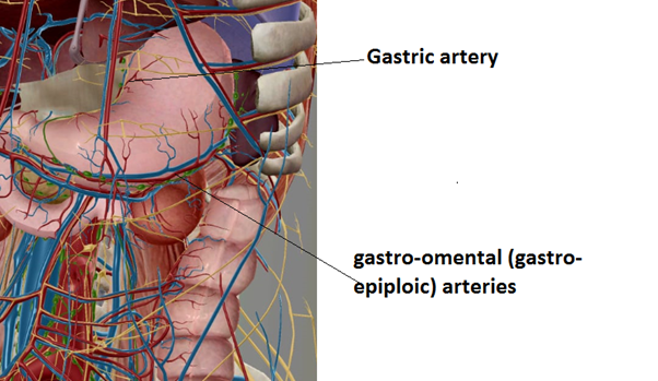
The stomach has the rich arterial supply derived from the celiac trunk, the first major visceral branch of the abdominal aorta.
- The left and right gastric arteries supply the lesser curvature of the stomach, which are branches of the celiac trunk and common hepatic artery, respectively.
- Left and right gastro-omental (gastro-epiploic) arteries supply the greater curvature, which is derived from the splenic and gastroduodenal arteries, respectively.
Stomach Anatomy, Lymphatic drainage:
The lymphatic drainage from the stomach passes through intermediary nodes before eventually reaching the celiac nodes. Gastric lymph drains into lymphatic vessels that arise in the mucosa, then create a rich network in the submucosa, and finally join together in a subperitoneal plexus.
Pathologies (Diseases):
| Gastric ulcers | Gastric ulcers are open sores on the stomach mucosa, while peptic ulcers are sores on the pyloric canal or, more frequently, the duodenum. The most common cause of stomach and duodenal ulcers is Helicobacter pylori (H. pylori). It is most common for people who experience chronic anxiety to develop peptic ulcers. Their gastric acid secretion rates are often markedly higher than normal between meals. The high acid in the stomach and duodenum is thought to overwhelm the bicarbonate normally as a result of sympathectomy. |
| Gastroesophageal reflux disease | Gastroesophageal reflux disease or GERD is a digestive disorder that affects the muscles that connect your esophagus to your stomach. This area is known as the lower esophageal sphincter (LES). A person suffering from this disease may get heartburn or acid indigestion. |
| Gastric cancer | In gastric cancer, malignant (cancer) cells form in the stomach lining. Gastric cancer risk is affected by age, diet, and stomach disease. Indigestion and abdominal discomfort or pain are signs of gastric cancer. |